Mitochondrial Biogenesis Protocols: PGC-1α Activation & Cellular Energy
Cellular energy capacity, systemic metabolic flexibility, and longevity are driven by the structural integrity and density of the mitochondrial pool. Peroxisome proliferator-activated receptor gamma coactivator 1-alpha (PGC-1α) acts as the master transcriptional coactivator responsible for mitochondrial biogenesis—the birth of new, highly efficient cellular powerhouses. Age-related mitochondrial decay, sedentary lifestyles, and nutrient excess suppress PGC-1α activity, driving energetic deficits, insulin resistance, and elevated systemic oxidative stress. Stimulating PGC-1α requires targeted metabolic stress and pharmacological modulation.
1. The AMPK / SIRT1 / PGC-1α Signaling Axis
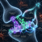
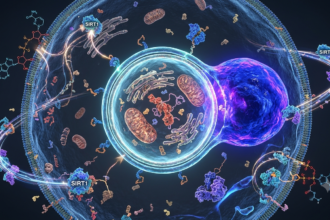
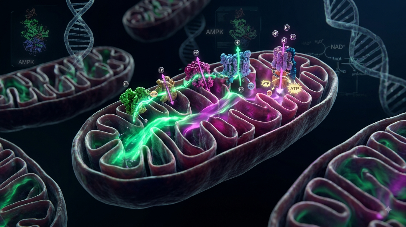
The activation of PGC-1α relies on two primary energy-sensing nodes: AMPK (5′ AMP-activated protein kinase) and SIRT1 (Sirtuin 1). When cellular ATP levels drop (reflected by high AMP/ATP ratios), AMPK directly phosphorylates PGC-1α. Simultaneously, elevated NAD+ levels activate SIRT1, which deacetylates PGC-1α, converting it into its active transcriptional state. Once activated, PGC-1α translocates to the nucleus to induce nuclear respiratory factors (NRF-1/NRF-2) and TFAM (mitochondrial transcription factor A), driving mitochondrial DNA replication.
2. Cellular Bioenergetic State Analysis
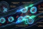
Accumulation of damaged, fragmented mitochondria coupled with suppressed biogenesis leads to chronic fatigue, low aerobic capacity, excess reactive oxygen species (ROS), and impaired cellular respiration.
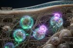
Robust expansion of mitochondrial volume fraction in muscular and neural tissues, maximizing ATP production, metabolic flexibility, and systemic physical/cognitive endurance.
3. Key Molecules for Biogenesis Activation
4. Environmental & Thermal Drivers of Biogenesis
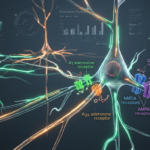
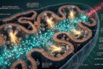
Exogenous nutraceuticals work synergistically with physical hormetic stressors. Acute cold exposure triggers cold-shock response mechanisms via norepinephrine release, activating beta-3 adrenergic receptors. This induces brown adipose tissue (BAT) thermogenesis and upregulates mitochondrial uncoupling protein 1 (UCP1) through powerful PGC-1α gene transcription. Combine this with endurance-based Zone 2 cardiovascular training to maximize mitochondrial volume and cristae density in skeletal muscle.
5. The Mitochondrial Expansion Protocol
Utilize intermittent fasting windows combined with NAD+ precursors and Urolithin A to purge senescent, inefficient mitochondria and elevate the intracellular NAD+/NADH ratio.
Engage in low-intensity steady-state (Zone 2) aerobic exercise supported by PQQ supplementation and cold therapy to activate the PGC-1α/TFAM cascade for new organelle synthesis.
6. Global Wellness Lab Verdict
Mitochondrial capacity is the ultimate rate-limiting factor in cellular energy output and lifespan. By strategically coupling targeted biogenic compounds like PQQ and NAD+ boosters with physical hormetic protocols, you systematically trigger PGC-1α upregulation—expanding your biological energy envelope.
- Puigserver, P., & Spiegelman, B. M. (2003). “Peroxisome proliferator-activated receptor-gamma coactivator 1 alpha (PGC-1alpha): transcriptional coactivator and metabolic regulator.” Endocrine Reviews, 24(1), 78-90.
- Chowanadisai, W., et al. (2010). “Pyrroloquinoline quinone stimulates mitochondrial biogenesis through cAMP response element-binding protein phosphorylation and increased PGC-1alpha expression.” Journal of Biological Chemistry, 285(1), 142-152.
- Ryu, D., et al. (2016). “Urolithin A induces mitophagy and prolongs lifespan in C. elegans and increases muscle function in rodents.” Nature Medicine, 22(8), 879-888.
- Cantó, C., et al. (2009). “AMPK regulates energy expenditure by modulating NAD+ metabolism and SIRT1 activity.” Nature, 458(7241), 1056-1060.
- Hood, D. A., et al. (2016). “Unraveling the mechanisms regulating muscle mitochondrial biogenesis.” Biochemical Journal, 473(9), 1059-1065.
Global Wellness Lab
“Resilience is not the absence of stress, but the biological capacity to manage it without systemic degradation.”
Page Contents
Toggle