Endothelial Shear Stress: Molecular Regulation of Nitric Oxide Synthesis and Vascular Glycocalyx Integrity
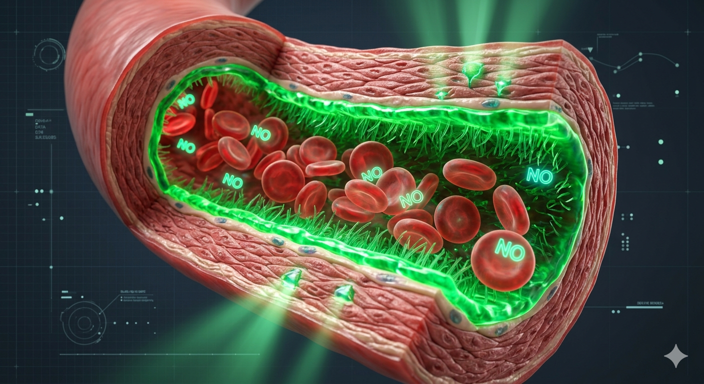
The human vascular endothelium is not merely a passive barrier separating the bloodstream from surrounding tissues; it operates as a dynamic, mechanosensitive endocrine organ. It continuously monitors the physical friction exerted by moving blood—a mechanical force quantified as endothelial shear stress (ESS).
When blood flow follows a steady, unidirectional laminar pattern, high ESS activates complex cellular structures that prompt the continuous release of protective signaling molecules. Conversely, when vascular branching or structural damage alters fluid dynamics into turbulent or oscillatory patterns, low ESS triggers an inflammatory cascade that degrades the blood vessel’s primary protective shield: the endothelial glycocalyx.
1. Mechanotransduction Pathways and eNOS Activation
Laminar blood flow passing over endothelial cells exerts a physical pulling force that deforms specialized mechanoreceptors on the cell surface. This mechanical movement activates the PI3K/Akt pathway, which directly leads to the phosphorylation of Endothelial Nitric Oxide Synthase (eNOS) at the specific amino acid site Serine 1177.
Active eNOS converts the amino acid L-arginine into nitric oxide (NO), a highly diffusible gas molecule. NO moves rapidly into the underlying vascular smooth muscle cells, activating soluble guanylyl cyclase (sGC) and increasing cyclic guanosine monophosphate (cGMP). This process decreases intracellular calcium concentrations, inducing vital, life-preserving vasodilation that lowers systemic blood pressure.
2. Hemodynamic Flow Profiles and Endothelial Responses
The physical pattern of blood flow dictates whether endothelial cells release protective or inflammatory signals. The table below outlines how specific flow characteristics alter vascular biology:
| Flow Profile Type | Shear Stress Level | Molecular/Cellular Consequence |
|---|---|---|
| Unidirectional Laminar | High Stable ESS | Upregulates eNOS, stabilizes Kruppel-like Factor 2 (KLF2), and actively suppresses cellular apoptosis. |
| Oscillatory / Disturbed | Low/Variable ESS | Triggers NADPH oxidase assembly, producing reactive oxygen species (ROS) that neutralize nitric oxide. |
| Turbulent Branching | Disrupted ESS | Upregulates adhesion molecules (VCAM-1 and ICAM-1), initiating leukocyte recruitment. |
3. Degradation of the Endothelial Glycocalyx Layer
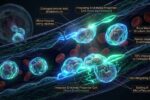
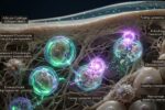
Sitting directly atop the endothelial cell lining is the glycocalyx—a delicate, gel-like network of glycosaminoglycans, proteoglycans, and bound plasma proteins. This structure acts as the vessel’s primary physical and chemical shield, preventing inflammatory white blood cells and low-density lipoproteins (LDL) from making direct contact with the vessel wall.
Under conditions of systemic oxidative stress or turbulent blood flow, activated enzymes known as metalloproteinases and heparanases begin shedding these delicate sugar chains. The loss of this gel layer leaves the artery walls exposed, allowing circulating fats to infiltrate the subendothelial space and accelerating the development of atherosclerotic plaques.
4. Micronutrient Substrates for Endothelial Structural Regeneration
Protecting the structural integrity of blood vessels involves providing specific substrates that limit oxidative shedding while supporting natural nitric oxide synthesis:
| Target System | Nutritional Cofactor | Biochemical Action |
|---|---|---|
| Glycocalyx Synthesis | Glucosamine & Hyaluronic Acid | Provides foundational building blocks used by endothelial cells to rebuild degraded glycosaminoglycan chains. |
| eNOS Uncoupling Prevention | L-Citrulline Malate | Acts as a highly bioavailable precursor that increases plasma L-arginine levels, ensuring optimal substrate availability for continuous NO synthesis. |
| Superoxide Neutralization | Coenzyme Q10 (Ubiquinol) | Suppresses vascular endothelial cell free-radical production, preserving newly synthesized nitric oxide gas molecules from oxidation. |
Clinical Focus: Asymmetric Dimethylarginine (ADMA)
Asymmetric dimethylarginine (ADMA) is an endogenous chemical compound that acts as a direct competitive inhibitor of eNOS. When tissue oxidative stress is elevated, the enzyme responsible for clearing ADMA degrades, causing it to pool within vessels. High circulating ADMA levels choke off healthy nitric oxide synthesis, making it a critical biomarker for assessing early vascular damage and long-term endothelial dysfunction.
Conclusion: Protecting the Vascular Architecture
Maintaining optimal cardiovascular function requires focus on both structural tissue integrity and complex fluid dynamics. The endothelium acts as a sensitive regulator, translating the mechanical friction of blood flow into vital chemical signals.
By providing targeted raw materials to support the endothelial glycocalyx and neutralizing chronic oxidative stress, we can support clean, laminar blood flow patterns, protecting deep vascular architecture and long-term heart health.
Global Wellness Lab
“Sustaining high endothelial shear stress and protecting glycocalyx thickness are structural prerequisites for avoiding systematic arterial degradation.”
2. Tarbell, J. M. et al., “The Endothelial Glycocalyx Layer: Mechanics of Protection and Shedding” – Cardiovascular Research, 2025.
This technical analysis outlines organic lipid architectures and must not replace custom clinical diagnosis. For advanced cognitive deficits, specialized electroencephalogram testing, or prescription adjustments, consult a certified neurologist.
*** This report is for technical educational purposes. Global Wellness Lab provides independent data for consumer safety and may receive commissions via certified affiliate links. ***
Page Contents
Toggle