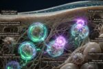

Macrophage Polarization and the NLRP3 Inflammasome: Molecular Switches Driving Immune Activation
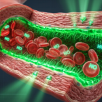
The innate immune system relies heavily on the functional plasticity of macrophages—highly versatile phagocytic cells tasked with tissue surveillance, pathogen elimination, and debris clearance. Rather than maintaining a fixed physiological state, macrophages continuously adapt their functional capabilities in response to microenvironmental cues.
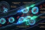
This adaptation process, known as macrophage polarization, primarily balances between two contrasting states: the pro-inflammatory M1 phenotype and the anti-inflammatory, pro-resolving M2 phenotype. When metabolic shifts or cellular damage alter this delicate immunological equilibrium, the aberrant assembly of a multi-protein intracellular sensor called the NLRP3 inflammasome can drive persistent, low-grade systemic tissue destruction.
1. The Immunometabolic Switch Behind M1 and M2 Phenotypes
When a macrophage detects lipopolysaccharides (LPS) or interferon-gamma (IFN-γ), it undergoes a profound metabolic remodeling process called the Warburg-like glycolytic switch. The cell shuts down its mitochondrial Krebs cycle at two specific points, leading to an accumulation of intracellular succinate.
This accumulated succinate stabilizes Hypoxia-Inducible Factor 1-Alpha (HIF-1α), which directly drives the expression of pro-inflammatory cytokines, solidifying the classic M1 state. Conversely, activation via interleukins like IL-4 promotes an intact Krebs cycle fueled by fatty acid oxidation, generating the oxidative phosphorylation required for tissue repair, extracellular matrix deposition, and anti-inflammatory signaling characteristic of the M2 state.
2. Immunological Disparities Between M1 and M2 States
The physiological function of a tissue macrophage depends entirely on its structural polarization vector. The table below details the molecular differences between these two states:
| Phenotypic Metric | M1 Classical Activation | M2 Alternative Activation |
|---|---|---|
| Primary Inducers | LPS, IFN-γ, Saturated FFAs | IL-4, IL-13, Glucocorticoids |
| Metabolic Pathway | Aerobic Glycolysis (Warburg) | Oxidative Phosphorylation & FAO |
| Arginine Metabolism | iNOS pathway (Produces Nitric Oxide) | Arg1 pathway (Produces Ornithine & Proline) |
| Primary Output | TNF-α, IL-6, IL-1β, ROS | IL-10, TGF-β, Collagen, Resolvins |
3. The NLRP3 Inflammasome Activation Cascade
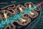
The hallmark of a chronically polarized M1 macrophage is its heightened vulnerability to assembling the NLRP3 inflammasome. This molecular complex requires a two-step licensing process. First, priming via surface receptors upregulates the genetic transcription of inactive NLRP3 and pro-IL-1β proteins.
The second triggering step involves physical stressors, such as extracellular ATP exposure, mitochondrial DNA leaking into the cytoplasm, or uric acid crystals piercing intracellular vesicles. These events trigger potassium efflux, prompting NLRP3 to link with ASC proteins and pro-caspase-1. The assembled complex then cleaves precursor signaling strands into mature, highly inflammatory IL-1β molecules, initiating localized cellular damage.
4. Micronutrient Regulators for Immunometabolic Polarization
Calming chronic M1 macrophage activity requires stabilizing mitochondrial structural boundaries and inhibiting downstream inflammasome assembly. Targeted compounds assist these biochemical mechanisms:
| Biochemical Pathway | Target Nutrient | Action on Macrophage Core |
|---|---|---|
| NLRP3 Assembly Blockade | Quercetin Monohydrate | Suppresses ASC oligomerization, directly interfering with the structural formation of the active inflammasome. |
| M2 Phenotype Support | Omega-3 Phospholipids (EPA) | Acts as a biological substrate for lipid mediators like resolvins, assisting the shift toward tissue-repairing M2 states. |
| Mitochondrial ROS Mitigation | Melatonin (Endogenous Pathway) | Scavenges reactive oxygen species inside the mitochondrial matrix, downregulating the signals that trigger NLRP3 assembly. |
Clinical Focus: Pyroptosis Induction
Unchecked activation of the caspase-1 pathway within an active NLRP3 complex can drive macrophages into a specialized form of inflammatory programmed cell death known as pyroptosis. During pyroptosis, the cell membrane ruptures violently, spilling large volumes of undiluted pro-inflammatory signaling proteins into the extracellular fluid. This cascade damages neighboring healthy tissues and perpetuates a state of chronic, low-grade systemic inflammation.
Conclusion: Rebalancing Immunometabolic Architecture
Resolving persistent immune stress requires looking beyond superficial symptoms to examine cell polarization mechanics. Macrophages demonstrate how tightly metabolic pathways are interwoven with our structural defenses.
By providing targeted compounds to suppress inflammasome assembly and neutralizing intracellular mitochondrial stressors, we can support the natural transition toward tissue-repairing immune states, protecting long-term systemic stability.
Global Wellness Lab
“Regulating macrophage polarization kinetics and limiting NLRP3 assembly are foundational requirements for controlling systematic tissue degradation.”
2. Schroder, K. et al., “The NLRP3 Inflammasome: Priming, Activation, and Pyroptotic Kinematics” – Cell Research, 2025.
This technical analysis outlines organic lipid architectures and must not replace custom clinical diagnosis. For advanced cognitive deficits, specialized electroencephalogram testing, or prescription adjustments, consult a certified neurologist.
*** This report is for technical educational purposes. Global Wellness Lab provides independent data for consumer safety and may receive commissions via certified affiliate links. ***
Page Contents
Toggle