Visceral Adipose Tissue Dysfunction: Molecular Mechanisms of Leptin Resistance and Lipotoxicity
In classical endocrinology, adipose tissue was long categorized as an inert storage depot for excess triglycerides. Modern metabolic biochemistry, however, has unveiled it as a highly sophisticated, active endocrine organ. The physiological profile changes completely when looking at subcutaneous fat versus visceral adipose tissue (VAT) surrounding internal organs.
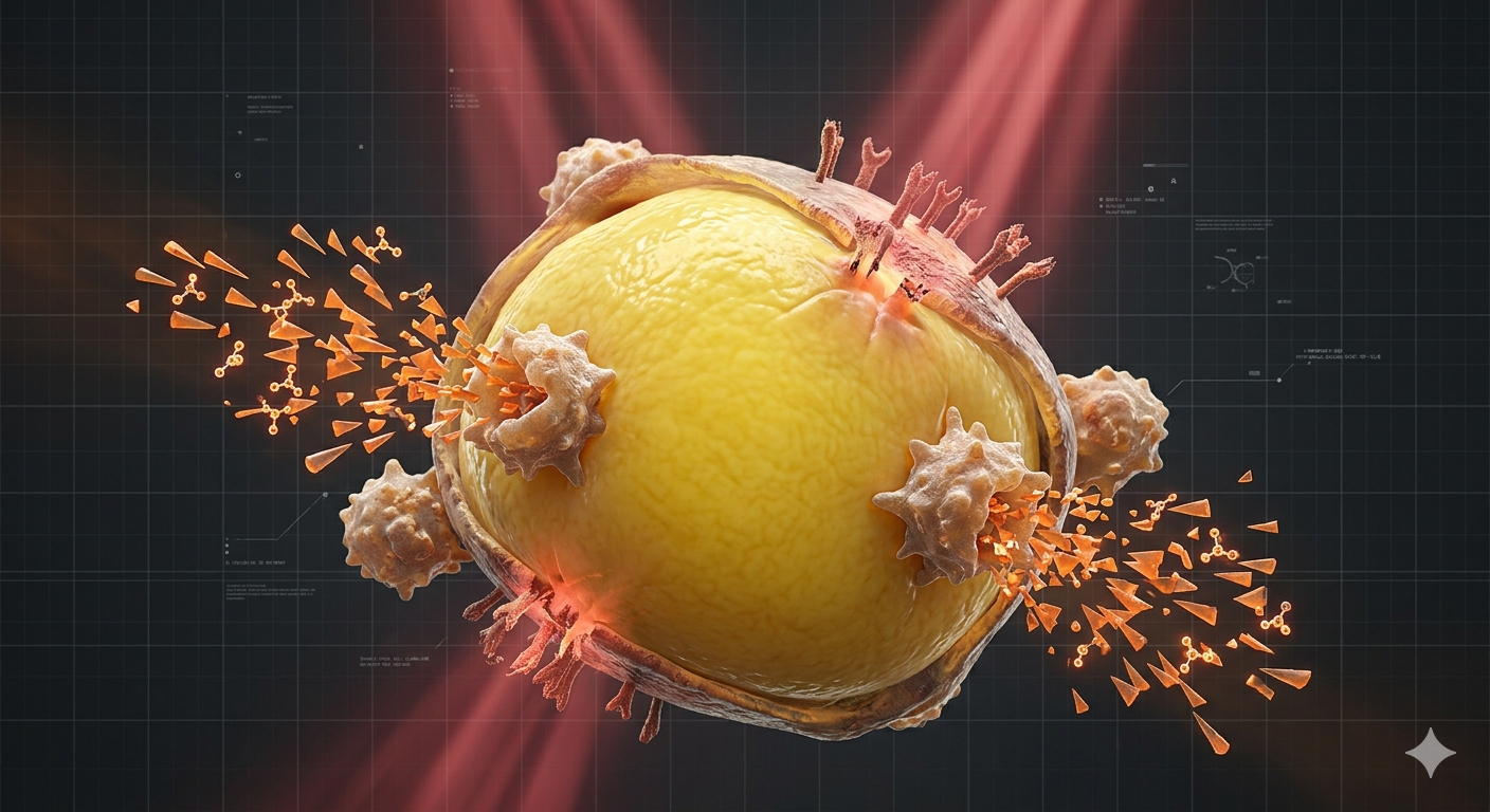
When chronic energy surplus forces visceral adipocytes past their physiological storage boundaries, the tissue enters a state of structural dysfunction. This hypertrophic expansion limits oxygen supply, setting off an aggressive wave of local tissue hypoxia, cellular necrosis, and systemic biochemical signaling degradation.
1. Adipocyte Hypertrophy, Hypoxia, and Macrophage Recruitment
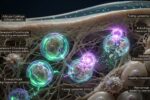
As visceral adipocytes expand to absorb lipids, they eventually exceed the diffusion capacity of local capillaries. The resulting intracellular hypoxia stabilizes Hypoxia-Inducible Factor 1-Alpha (HIF-1α). This transcription factor initiates a defensive but destructive cascade, upregulating extracellular matrix collagen cross-linking and inducing mechanical tissue fibrosis.
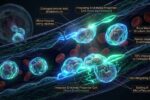
Stressed, hypoxic adipocytes begin to rupture, leaking free fatty acids and cellular debris into the surrounding tissue. This cellular emergency alters the local immune profile, shifting resting M2 macrophages into pro-inflammatory M1 phenotypes. These immune cells cluster around dead fat cells in “crown-like structures,” releasing large amounts of destructive cytokines.
2. The Shift in Adipokine Secretion Profiles
Healthy adipose tissue releases protective compounds like adiponectin to preserve insulin sensitivity. In a dysfunctional visceral state, this secretion balance flips completely, shifting toward markers that promote systemic insulin resistance and cardiovascular strain:
| Biochemical Marker | Secretory Trend in VAT | Primary Target Response |
|---|---|---|
| Adiponectin | Severely Suppressed | Reduces fatty acid oxidation in muscle tissue and downregulates liver AMPK pathways. |
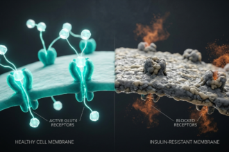
| Tumor Necrosis Factor (TNF-α) | Significantly Elevated | Triggers serine phosphorylation of IRS-1, directly short-circuiting insulin receptor signaling. |
| Interleukin-6 (IL-6) | Significantly Elevated | Travels via portal circulation to the liver, accelerating C-Reactive Protein (CRP) synthesis. |
| Leptin | Chronically Elevated | Saturates blood-brain barrier transport mechanisms, inducing deep central satiety resistance. |
3. The Central Mechanism of Leptin Resistance
Leptin acts as the master hormonal signal for energy abundance, released by fat cells to tell the hypothalamus to reduce appetite and increase energy expenditure. Under conditions of chronic visceral hypertrophy, excessive leptin production floods the system, desensitizing long-form leptin receptors (Ob-Rb) in the brain.
This disruption happens through the upregulation of SOCS3 (Suppressor of Cytokine Signaling 3). SOCS3 binds directly to the JAK2 tyrosine kinase complex, halting the STAT3 phosphorylation cascade. The brain becomes completely blind to body fat signals, misinterpreting the excess energy state as starvation and driving continuous hunger.
4. Targeted Interventions for Adipose Recalibration
Reversing visceral tissue dysfunction requires reducing local inflammation and restoring blood-brain barrier leptin transport kinetics. Specialized natural compounds support these biochemical pathways:
| Target Pathway | Nutritional Catalyst | Biochemical Action |
|---|---|---|
| NF-kB Downregulation | Curcumin C3 Complex | Blocks the IκB kinase complex, reducing macrophage transcription of TNF-α and IL-6. |
| SOCS3 Suppression | EGCG (Green Tea Extract) | Lowers intracellular SOCS3 expression, helping restore central hypothalamic JAK2-STAT3 signaling. |
| Lipophilicity Regulation | Alpha-Lipoic Acid (R-ALA) | Supports GLUT4 migration and acts as a cofactor for mitochondrial pyruvate dehydrogenase. |
Clinical Focus: Free Fatty Acid (FFA) Spillover
When visceral adipose storage capacity is completely overwhelmed, fat spills over into non-adipose organs. This ectopic fat deposition targets the liver and pancreas, triggering local insulin resistance and beta-cell apoptosis. Managing visceral health requires focus on adipocyte hypertrophy, lipid clearance kinetics, and chronic low-grade systemic inflammation.
Conclusion: Shifting From Lipotoxicity to Adipose Homeostasis
Resolving metabolic dysfunction requires looking beyond total body weight to examine specific tissue health. Visceral adipose tissue expansion drives systemic inflammation and central metabolic resistance across the human organism.
Combining specific nutrient strategies with targeted substrate clearance protocols calms chronic tissue inflammation, helping restore proper leptin signaling and long-term metabolic health.
Global Wellness Lab
“Ameliorating visceral fat lipotoxicity through targeted cellular interventions is a prerequisite for systemic inflammatory control.”
2. Friedman, J. M. et al., “Hypothalamic Leptin Resistance: SOCS3 and JAK2 Signaling Dynamics” – Cell Metabolism Reviews, 2025.
This technical analysis outlines organic lipid architectures and must not replace custom clinical diagnosis. For advanced cognitive deficits, specialized electroencephalogram testing, or prescription adjustments, consult a certified neurologist.
*** This report is for technical educational purposes. Global Wellness Lab provides independent data for consumer safety and may receive commissions via certified affiliate links. ***
Page Contents
Toggle