Autophagy Induction Protocols: mTOR Suppression & Lysosomal Recycling
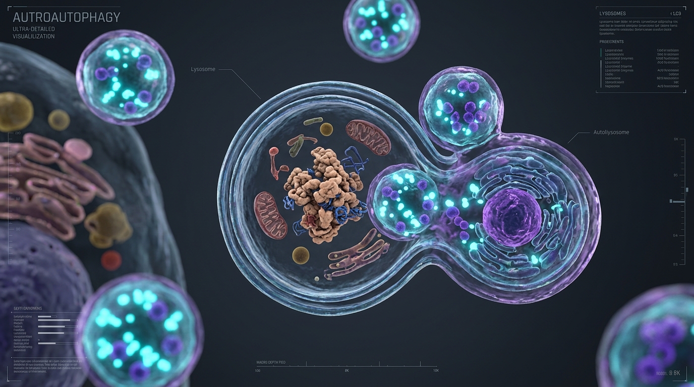
Autophagy is the fundamental, evolutionarily conserved lysosomal degradation pathway responsible for clearing protein aggregates, damaged organelles, and intracellular pathogens. In aging tissues, autophagic flux declines sharply due to persistent activation of the Mechanistic Target of Rapamycin Complex 1 (mTORC1). This reduction in autophagic capacity leads to proteostatic collapse, cellular senescence, and chronic metabolic dysfunction. Strategically downregulating mTORC1 while activating the ULK1/Atg1 complex restores lysosomal degradation and recycles cellular components into fresh amino acids and lipid substrates.
1. The mTORC1-ULK1 Signaling Axis
Under nutrient-replete conditions, active mTORC1 phosphorylates ULK1 at Ser 757, preventing its interaction with AMPK and suppressing phagophore initiation. Upon nutrient deprivation, amino acid withdrawal, or pharmacological mTOR inhibition, mTORC1 dissociates from the ULK1 complex. AMPK then directly phosphorylates ULK1 at Ser 317 and Ser 777, triggering downstream nucleation of the Beclin-1/Vps34 complex. This cascade synthesizes phosphatidylinositol 3-phosphate (PI3P) at the endoplasmic reticulum, initiating autophagosome formation.
2. Proteostatic State Analysis
Continuous nutrient sensing blocks autophagosome biogenesis, accumulating lipofuscin granules, damaged mitochondria, and triggering pro-inflammatory cellular signaling.
Pulsed mTOR suppression restores lysosomal acidic pH, increases LC3-II turnover, and recycles damaged proteins to maintain tissue metabolic youth.
3. Key Autophagy-Inducing Modulators
4. Transcriptional Control via TFEB Master Regulator
Transcription Factor EB (TFEB) controls the biogenesis of autophagosomes and lysosomes. Under normal nutrient conditions, TFEB is phosphorylated by mTORC1 on the lysosomal membrane and retained in the cytoplasm. When mTORC1 is inhibited or cellular stress occurs, unphosphorylated TFEB translocates directly to the cell nucleus. There, it upregulates the CLEAR gene network, accelerating vesicle maturation, lysosomal acid hydrolase synthesis, and the fusion rate between autophagosomes and lysosomes.
5. The Autophagic Clearance Protocol
Implement a 24-to-36 hour water fast every 14 days to naturally deplete liver glycogen, suppress serum insulin/IGF-1 signaling, and shut down mTORC1 activation in target peripheral tissues.
Supplement daily with high-purity Spermidine and Trehalose during non-fasting windows to maintain steady-state EP300 inhibition and stimulate continuous background proteostasis.
6. Global Wellness Lab Verdict
Long-term cellular health is unachievable without efficient autophagic clearance. By balancing nutrient sensing through pulsed mTORC1 downregulation and targeted activation of the TFEB pathway, cells can purge toxic protein debris and preserve functional integrity against biological aging.
- Mizushima, N., & Komatsu, M. (2011). “Autophagy: renovation of cells and tissues.” Cell, 147(4), 728-741.
- Madeo, F., et al. (2018). “Spermidine in health and disease.” Science, 359(6374), eaan2788.
- Settembre, C., et al. (2011). “TFEB links autophagy to lysosomal biogenesis.” Science, 332(6036), 1429-1433.
- Kim, J., et al. (2011). “AMPK and mTOR regulate autophagy through direct phosphorylation of Ulk1.” Nature Cell Biology, 13(2), 132-141.
- Harrison, D. E., et al. (2009). “Rapamycin fed late in life extends lifespan in genetically heterogeneous mice.” Nature, 460(7253), 392-395.
Global Wellness Lab
“Resilience is not the absence of stress, but the biological capacity to manage it without systemic degradation.”
Page Contents
Toggle